Özet
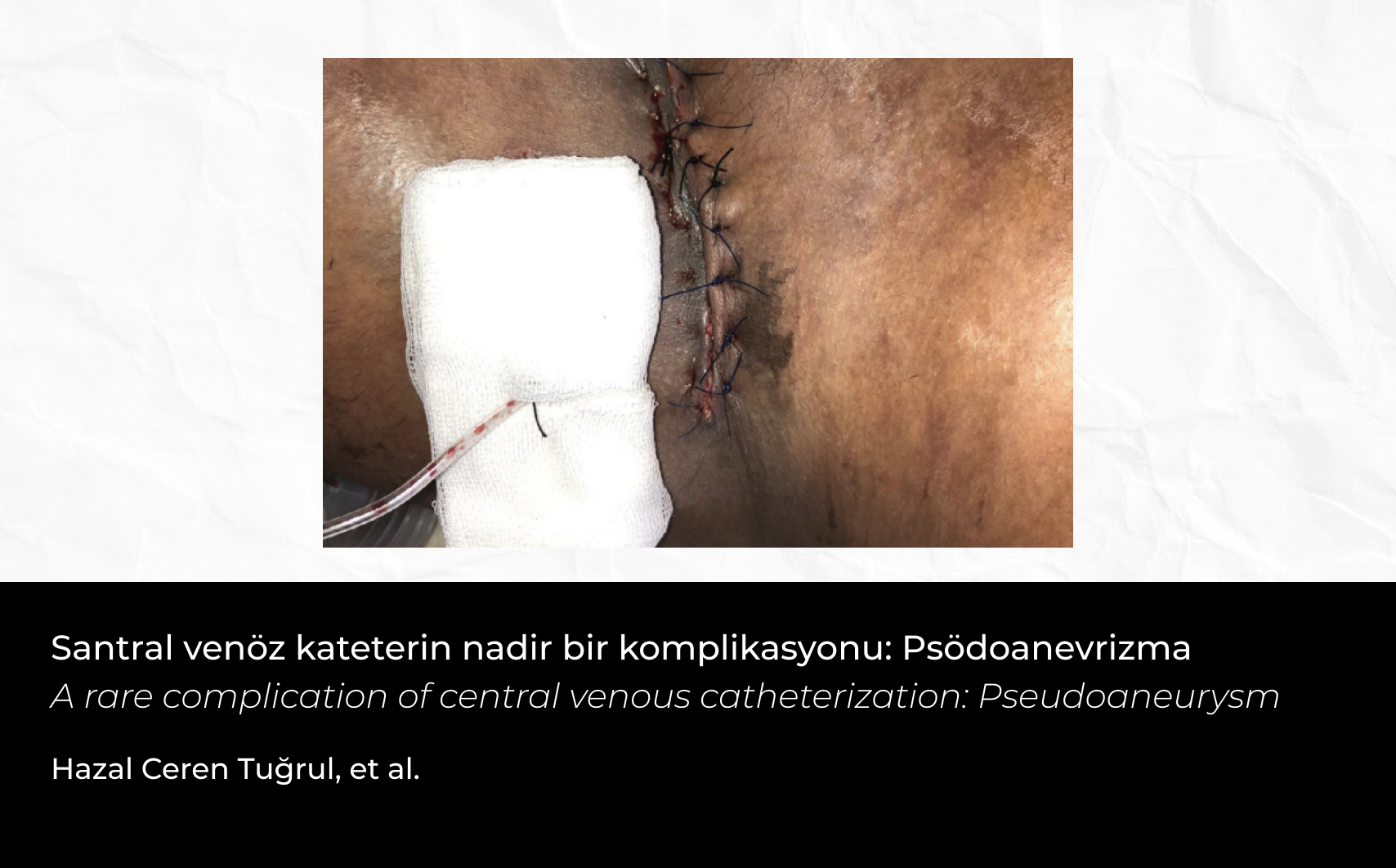
Ekstravazasyon, intravenöz olarak verilen vezikant maddelerin damar dışına sızarak çevre dokularda hasara yol açmasıdır. Özellikle küçük çocuklar ve iletişim sorunu yaşayan hastalar bu duruma karşı savunmasızdır. Ekstravazasyon, ciddi doku hasarına, uzun hastane yatışlarına ve maliyet artışına neden olabilir. Bu olgu sunumunda, hipertonik salin infüzyonu sonrası ciddi doku kaybı gelişen ve greft uygulanan 16 yaşındaki bir erkek hasta sunulmuştur. Hasta, trafik kazası sonrası subaraknoid kanama, pnömotoraks ve humerus kırığı tanılarıyla yoğun bakıma alınmış, mekanik ventilasyon altında tedavi edilmiştir. Hipertonik salin infüzyonu sonrası sol alt ekstremitede şişlik ve kızarıklık gözlenmiş, damaryolu çıkarılarak bölgeye soğuk uygulama yapılmıştır. Takipte nekrotik yara gelişmiş, yara bakım ekibi tarafından debritman yapılmış ve sekonder iyileşme hedeflenmiştir. Ancak yeterli iyileşme sağlanamayınca, split thickness deri grefti uygulanmış ve postoperatif 15. günde yara tamamen iyileşmiştir. Yoğun bakım ihtiyacı kalmayan hasta, pediatri servisine devredilmiştir.
Abstract
Extravasation is the leakage of vesicant substances administered intravenously into surrounding tissues, causing damage. Children and patients with communication difficulties are particularly vulnerable to this condition. Extravasation can lead to serious tissue damage, prolonged hospital stays, and increased healthcare costs. In this case report, we present a 16-year-old male patient who developed significant tissue loss following hypertonic saline infusion and subsequently required grafting. The patient was admitted to the intensive care unit after a traffic accident with diagnoses of subarachnoid hemorrhage, pneumothorax, and humeral shaft fracture. He was treated under mechanical ventilation. After the administration of hypertonic saline infusion, swelling and redness were observed in the left lower extremity, leading to the removal of the intravenous line and the application of cold therapy to the affected area. In follow-up, a necrotic wound developed, and the wound care team performed debridement with the aim of secondary healing. However, as secondary healing was insufficient, a split-thickness skin graft was applied. On postoperative day 15, the wound was completely healed. The patient, no longer requiring intensive care, was transferred to the pediatric ward.


 Hazal Ceren Tuğrul1


